
Introduction
RNase P is one of the most fascinating molecules in cell biology, and yet most people have never heard of it. RNase P (short for Ribonuclease P) is an enzyme found in every living cell — from the simplest bacteria to human beings — and it performs a job that no other enzyme can replace. Without RNase P, our cells could not make functional transfer RNA (tRNA), and without functional tRNA, protein synthesis would grind to a halt.
What makes RNase P truly remarkable is that, in most organisms, it is not a normal protein enzyme at all. It is a ribozyme, meaning the catalytic (cutting) work is done by RNA rather than protein. This discovery, made in the early 1980s, changed the way scientists thought about the origins of life and earned a Nobel Prize.
History
The story of RNase P is closely tied to one of the biggest breakthroughs in molecular biology.
- 1970s: Scientists first identified an enzymatic activity in bacterial cells (particularly Escherichia coli) responsible for cutting pre-tRNA molecules at a specific site.
- 1978: Sidney Altman and his research team demonstrated that this activity, later named RNase P, required both an RNA component and a protein component to function.
- 1983: Altman, together with Norman Pace and colleagues, made the groundbreaking discovery that the RNA component of RNase P — not the protein — was the actual catalytic part of the enzyme. This was one of the first demonstrations that RNA molecules could act as biological catalysts, a concept that was almost unthinkable at the time because scientists believed only proteins could catalyze reactions.
- In 1989, Sidney Altman and Thomas Cech were jointly awarded the Nobel Prize in Chemistry for their groundbreaking discovery that certain RNA molecules possess catalytic activity. These catalytic RNA molecules are commonly known as ribozymes.
- Later decades: Researchers discovered that RNase P exists in nearly all domains of life, including bacteria, archaea, eukaryotes (plants, fungi, animals, humans), and even some organelles like mitochondria and chloroplasts. In humans and some archaea, additional protein subunits were found to be essential alongside the catalytic RNA.
This history matters because it reshaped biology’s understanding of the “RNA World” hypothesis — the idea that early life may have relied on RNA molecules to both store genetic information and catalyze chemical reactions, before protein enzymes and DNA became dominant.
Classification
It can be classified in a few different ways, based on structure, catalytic component, and the organism in which it is found.
By Catalytic Component
- RNA-based RNase P (ribozyme form) – Found in bacteria, archaea, and many eukaryotic organelles. The RNA subunit itself carries out the chemical cutting reaction.
- Protein-only RNase P (PRORP) – Found in the nucleus and organelles of some eukaryotes, such as plants and certain protozoa. Here, the enzyme relies entirely on protein for catalysis, without any RNA component at all — a striking example of convergent evolution, where two very different molecular strategies evolved to solve the same biological problem.
By Organism Type
Organism Type | RNase P Composition | Example |
Bacteria | One RNA subunit + one small protein subunit | E. coli |
Archaea | One RNA subunit + multiple (usually 4–5) protein subunits | Methanothermobacter |
Eukaryotes (Nuclear) | One RNA subunit + up to 9–10 protein subunits | Human nuclear RNase P |
Human Mitochondria | Protein-only (PRORP-type in some species) or RNA-based in others | Mitochondrial RNase P |
Plants | Protein-only PRORP enzymes | Arabidopsis |
Types
Broadly, RNase P is grouped into the following functional types based on cellular location and molecular makeup:
- Bacterial-type RNase P – Simplest form, with one catalytic RNA and one protein subunit that stabilizes the RNA and helps it bind substrate.
- Archaeal-type RNase P – More complex, with several small protein subunits assisting the RNA core.
- Eukaryotic Nuclear RNase P – The most protein-rich version, found in the nucleus of human and other eukaryotic cells, with the RNA component still central to catalysis.
- Organellar RNase P – Found inside mitochondria and chloroplasts, sometimes RNA-based and sometimes entirely protein-based (PRORP), depending on the species.
- Protein-Only RNase P (PRORP) – A completely different family of enzymes that perform the same tRNA-processing job without using any RNA for catalysis.
Components and Parts
Understanding the “parts list” of RNase P helps explain how it works.
1. The RNA Subunit
- Also called the RNase P RNA (RPR).
- This is the catalytic heart of the enzyme in most organisms.
- It folds into a complex three-dimensional shape with two major structural domains:
- The Specificity (S) domain – helps recognize and bind the correct pre-tRNA substrate.
- The Catalytic (C) domain – contains the active site where the actual cutting reaction happens.
2. The Protein Subunit(s)
- In bacteria, there is just one small protein subunit.
- In archaea and humans, there are multiple protein subunits (in humans, at least nine have been identified).
- These proteins do not usually perform the chemical cutting themselves; instead, they:
- Stabilize the RNA’s folded structure.
- Improve binding to the substrate (pre-tRNA).
- Help the enzyme function properly under normal cellular conditions (protein-free RNA alone often needs unnaturally high salt concentrations to fold and work in a test tube).
3. The Substrate: Pre-tRNA
- The molecule RNase P acts upon is precursor tRNA (pre-tRNA), which has an extra “leader” sequence at its 5′ end that must be removed before the tRNA can function properly.
Principle
The basic principle behind RNase P’s action is site-specific endonucleolytic cleavage. This means the enzyme recognizes a very specific location on its RNA substrate and makes a single, precise cut, rather than randomly chopping up RNA molecules. This precision is essential because a wrong cut would produce a non-functional tRNA molecule, which could disrupt protein synthesis in the cell.
RNase P recognizes the overall three-dimensional shape (or “fold”) of pre-tRNA — particularly the cloverleaf-like secondary structure — rather than reading a specific sequence of letters. This is why RNase P can process many different tRNA molecules, even though their sequences vary, as long as they share the same general shape.
Working Principle and Mechanism
Here is a simplified, step-by-step explanation of how RNase P processes pre-tRNA:
- Substrate Recognition: The pre-tRNA molecule folds into its characteristic cloverleaf and L-shaped 3D structure. RNase P’s specificity domain recognizes this shape.
- Binding: RNase P binds to the pre-tRNA, correctly positioning the 5′ leader sequence within its catalytic core.
- Metal Ion Assistance: Magnesium ions (Mg²⁺) play a critical role. The catalytic RNA core coordinates these metal ions to help stabilize the transition state of the chemical reaction and position water molecules for the cutting step.
- Catalysis (Cleavage): A water molecule, activated with the help of magnesium ions, attacks the phosphate backbone at a specific bond, cleaving the RNA chain and releasing the 5′ leader sequence.
- Product Release: The mature tRNA (now missing its leader sequence) is released, ready for further processing steps, such as the addition of a CCA sequence at its 3′ end and various chemical modifications.
- Enzyme Recycling: RNase P is not consumed in the reaction — like all true enzymes and catalysts, it is released unchanged and can process another pre-tRNA molecule.
This entire process typically happens very quickly and is repeated constantly in every living cell, since tRNA processing is required continuously to keep protein synthesis running smoothly.
Working Principle and Mechanism
Here is a simplified, step-by-step explanation of how RNase P processes pre-tRNA:
- Substrate Recognition: The pre-tRNA molecule folds into its characteristic cloverleaf and L-shaped 3D structure. RNase P’s specificity domain recognizes this shape.
- Binding: RNase P binds to the pre-tRNA, correctly positioning the 5′ leader sequence within its catalytic core.
- Metal Ion Assistance: Magnesium ions (Mg²⁺) play a critical role. The catalytic RNA core coordinates these metal ions to help stabilize the transition state of the chemical reaction and position water molecules for the cutting step.
- Catalysis (Cleavage): A water molecule, activated with the help of magnesium ions, attacks the phosphate backbone at a specific bond, cleaving the RNA chain and releasing the 5′ leader sequence.
- Product Release: The mature tRNA (now missing its leader sequence) is released, ready for further processing steps, such as the addition of a CCA sequence at its 3′ end and various chemical modifications.
- Enzyme Recycling: RNase P is not consumed in the reaction — like all true enzymes and catalysts, it is released unchanged and can process another pre-tRNA molecule.
This entire process typically happens very quickly and is repeated constantly in every living cell, since tRNA processing is required continuously to keep protein synthesis running smoothly.
Applications
It is important well beyond basic biology. Its applications include:
1. Fundamental Molecular Biology Research
- Used as a model system for studying RNA catalysis, RNA folding, and enzyme evolution.
2. Understanding the RNA World Hypothesis
- It is one of the strongest pieces of evidence supporting the theory that early life used RNA molecules for both genetic storage and catalysis before DNA and proteins took over these roles.
3. Molecular Diagnostics (Clinical Laboratory Use)
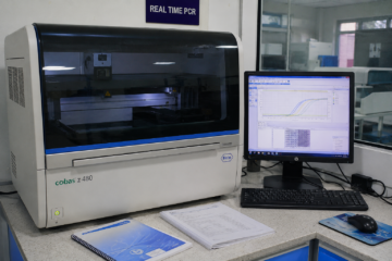
- The human RNase P gene (RPP30) is widely used as an internal control in real-time PCR (polymerase chain reaction) tests, including many SARS-CoV-2 (COVID-19) diagnostic assays. Since this gene is present in nearly every human cell, its detection confirms that a sample was collected properly and that the PCR reaction itself is working correctly.
4. Antibacterial Drug Development
- Because bacterial RNase P differs structurally from the human version, researchers are exploring it as a potential target for new antibiotics that would not harm human cells.
5. Biotechnology and Gene-Targeting Tools
- Modified, engineered versions of RNase P (called “external guide sequence” or EGS technology) have been studied as tools to cut and potentially silence specific target RNA molecules, including viral RNA, in experimental research settings.
Advantages
RNase P offers several biological and scientific advantages:
- High specificity: Cuts only at the correct site on pre-tRNA, preventing errors in protein synthesis machinery.
- Universality: Present in nearly all life forms, making it an excellent comparative tool for evolutionary studies.
- Reusability: As a true catalyst, one RNase P molecule can process many substrate molecules repeatedly.
- Diagnostic reliability: The human RNase P gene assay provides a dependable internal quality control for molecular tests, reducing false-negative results caused by poor sample collection.
- Research value: Serves as a powerful model for studying ribozymes, informing broader research into RNA-based therapeutics.
Disadvantages / Limitations
Like any biological system, RNase P also comes with certain limitations:
- Complexity of eukaryotic forms: Human RNase P has many protein subunits, making it harder to study and reconstruct in a laboratory setting compared to the simpler bacterial version.
- Difficulty in isolating catalytic RNA alone: In many organisms, the RNA component alone is unstable or inefficient without its protein partners, complicating purification for research.
- Limited direct clinical treatment applications so far: While RNase P-based technologies (like EGS) show promise, they are still largely experimental and not yet widely used as approved therapies.
- Species variability: Differences between bacterial, archaeal, and eukaryotic RNase P structures mean that findings from one organism cannot always be directly applied to another.
Clinical Significance
RNase P has several important connections to human health and clinical laboratory science:
1. Essential for Normal Cell Function
Since every human cell needs functional tRNA to build proteins, RNase P activity is essential for basic cellular survival. Severe disruption of RNase P function would be incompatible with normal cell growth, which is why mutations affecting core RNase P components are extremely rare and, when they occur in certain subunits, have been linked to specific inherited conditions affecting bone marrow function and tissue development in some research studies.
2. Use in Infectious Disease Testing
The human RNase P (RP) gene is one of the most widely used internal controls in molecular diagnostic testing, especially in real-time reverse transcription PCR (RT-PCR) assays for detecting viruses, including SARS-CoV-2. Detecting the human RNase P gene in a patient sample confirms:
- Adequate sample collection (enough human cellular material was obtained).
- Proper RNA/DNA extraction.
- Correct functioning of the PCR reaction itself.
3. Target for Future Antimicrobial Therapy
Because the RNA-based catalytic mechanism in bacteria differs from the human system, some researchers view bacterial RNase P as a potential future target for novel antibiotics, especially important given rising antibiotic resistance worldwide.
4. Contribution to Basic Understanding of Genetic Disease
Research into RNase P processing errors contributes to a broader understanding of how disruptions in RNA processing pathways can lead to cellular dysfunction, informing research into various genetic and hematological disorders.
Interpretation of Results (In Diagnostic Testing Context)
When RNase P is used as an internal control gene in a diagnostic RT-PCR test (such as certain COVID-19 assays), interpretation typically follows this pattern:
RNase P (Internal Control) Result | Target Pathogen Result | Interpretation |
Positive (detected) | Positive | Valid test; pathogen detected |
Positive (detected) | Negative | Valid test; pathogen not detected (true negative) |
Negative (not detected) | Negative | Invalid test; poor sample quality or collection error — repeat testing required |
Negative (not detected) | Positive | Uncommon; may still be considered valid depending on the specific assay protocol, but often flagged for review |
In simple terms: if RNase P is not detected at all, it usually means the sample did not contain enough human cellular material, and the test result cannot be trusted — a new sample should be collected and tested again.
Frequently Observed Errors
In laboratory settings using RNase P as an internal control, some common errors include:
- Inadequate sample collection – Swabs that do not collect enough cellular material, leading to a falsely negative RNase P signal.
- RNA degradation – Poor sample storage or transport conditions causing RNA breakdown before testing.
- Pipetting errors – Inaccurate volumes of reagents or samples affecting PCR amplification.
- Contamination – Cross-contamination between samples leading to false positive results.
- Reagent expiration – Using expired primers, probes, or master mix reducing assay sensitivity.
- Thermal cycler miscalibration – Incorrect temperature cycling affecting amplification efficiency.
Troubleshooting
If the RNase P internal control fails to amplify (is undetected) during PCR testing, laboratory staff typically follow these troubleshooting steps:
- Check sample collection documentation – Confirm the swab or sample type met collection guidelines.
- Re-extract RNA/DNA – Repeat the nucleic acid extraction step in case of technical failure.
- Verify reagent integrity – Confirm primers, probes, and master mix have not expired or been improperly stored.
- Inspect equipment calibration – Ensure the thermal cycler is functioning within specifications.
- Repeat the test with a fresh sample – If available, request a new sample from the patient.
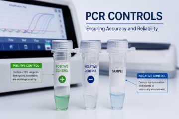
- Review positive and negative controls – Confirm that the overall PCR run controls behaved as expected, ruling out a systemic run failure versus a single-sample issue.
Care and Maintenance (Laboratory Reagents and Equipment)
Although RNase P is a naturally occurring biological molecule rather than laboratory equipment, the reagents used in RNase P-based testing—such as primers, probes, and enzyme mixes—must be carefully handled, stored, and prepared to maintain their stability, prevent contamination, and ensure accurate and reliable diagnostic results:
Store PCR reagents at the manufacturer-recommended temperature (commonly -20°C for master mixes and primers).
- Avoid repeated freeze-thaw cycles, which can degrade RNA-based reagents and reduce assay sensitivity.
- Use RNase-free consumables (tubes, tips, water) throughout the workflow, since environmental RNases can degrade both samples and control RNA.
- Regularly calibrate thermal cyclers and pipettes according to laboratory quality standards.
- Label and date all reagents clearly, discarding expired materials promptly.
Quality Assurance (IQC and EQA)
Quality assurance is critical when RNase P is used in molecular diagnostics.
Internal Quality Control (IQC)
- Every diagnostic PCR run should include the human RNase P gene as an internal control for each patient sample, alongside separate positive and negative controls for the overall run.
- IQC ensures that each individual sample was collected, processed, and amplified correctly.
External Quality Assessment (EQA)
- Laboratories participate in external proficiency testing programs, where blinded samples are sent by an accredited body to check that a laboratory’s testing process (including RNase P internal control performance) meets required standards.
- EQA helps confirm inter-laboratory consistency and supports accreditation processes (such as those following CLSI — Clinical and Laboratory Standards Institute — guidelines).
Safety Precautions
When working with RNase P-related reagents and clinical samples in a laboratory setting, standard biosafety practices apply:
- Wear appropriate personal protective equipment (PPE), including gloves, lab coats, and eye protection.
- Handle all patient samples as potentially infectious, following standard biosafety level protocols relevant to the pathogen being tested.
- Dispose of biological waste and used reagents according to institutional and regulatory biohazard waste guidelines.
- Avoid mouth pipetting and always use mechanical pipetting devices.
- Decontaminate work surfaces regularly with appropriate disinfectants to prevent RNase contamination, which can degrade RNA samples and affect test accuracy.
Key Points
- It is a ribonucleoprotein enzyme responsible for processing precursor tRNA into mature, functional tRNA.
- In most organisms, RNase P is a ribozyme, meaning its RNA component — not its protein component — carries out the catalytic cutting reaction.
- The discovery of RNase P’s catalytic RNA in the early 1980s helped establish the “RNA World” hypothesis and earned Sidney Altman a share of the 1989 Nobel Prize in Chemistry.
- RNase P is essential for protein synthesis in all cells, since functional tRNA is required to build proteins.
- The human RNase P gene is widely used as an internal control in molecular diagnostic PCR tests, including several SARS-CoV-2 detection assays.
- Some eukaryotic organisms, like plants, use a completely different, protein-only version of RNase P called PRORP.
- Understanding RNase P has research applications ranging from evolutionary biology to potential future antibiotic development.
Conclusion
RNase P may be microscopic and largely unknown outside scientific circles, but its role in biology is enormous. As one of the first discovered examples of a ribozyme — an RNA molecule capable of acting as a true enzyme — RNase P helped reshape our understanding of how life may have originated and how cells continue to function today. Its essential job of converting precursor tRNA into mature, usable tRNA keeps the protein-making machinery of every cell running smoothly, from simple bacteria to complex human tissues.
Beyond its foundational biological role, RNase P has found a very practical place in modern medicine: as a reliable internal control gene in molecular diagnostic testing, including many of the PCR-based tests used during the COVID-19 pandemic. This dual identity — as both an ancient evolutionary clue and a modern diagnostic tool — makes RNase P a wonderful example of how basic scientific research, sometimes decades old, can quietly support real-world healthcare applications.
Whether you are a student memorizing enzyme functions for an exam, a laboratory professional troubleshooting a failed PCR run, or simply someone curious about the hidden machinery of life, understanding RNase P offers a deeper appreciation for the elegant and efficient systems that keep every living cell functioning correctly.
Frequently Asked Questions (FAQs)
What is RNase P in simple terms?
RNase P is an enzyme found in nearly all living cells that trims off an extra piece of RNA from newly made transfer RNA (tRNA), turning it into its mature, functional form.
Is RNase P a protein or an RNA molecule?
In most organisms, RNase P is made of both RNA and protein, but the RNA component does the actual cutting work, making RNase P a “ribozyme.” Some organisms, like plants, have a protein-only version instead.
Why is RNase P important in COVID-19 testing?
The human RNase P gene is used as an internal control in many COVID-19 PCR tests. Detecting it confirms that a sample was collected properly and that the test itself is working correctly, helping to avoid false-negative results.
What happens if RNase P does not work properly?
Since RNase P is essential for producing functional tRNA, severe disruption of its activity would impair protein synthesis, which is incompatible with normal cell survival — this is one reason mutations severely affecting core RNase P function are extremely rare.
Where is RNase P found in the body?
RNase P is found in essentially every human cell, since all cells need functional tRNA to produce proteins. It is located primarily in the cell nucleus, though related versions also exist in mitochondria.
Who discovered RNase P's catalytic ability?
Sidney Altman and colleagues, including Norman Pace, discovered in 1983 that the RNA component of RNase P — not the protein — was responsible for its catalytic activity, a finding that contributed to Altman receiving the 1989 Nobel Prize in Chemistry.
Is RNase P the same in bacteria and humans?
No. While the core function is similar, bacterial RNase P is simpler, with just one small protein subunit, whereas human RNase P has multiple protein subunits (up to nine or more) supporting its RNA core.
Can RNase P be used to develop new medicines?
Researchers are exploring bacterial RNase P as a potential target for new antibiotics, and engineered versions of RNase P (external guide sequence technology) are being studied experimentally as tools to target specific RNA molecules, though these applications remain largely in the research stage.
What is the difference between RNase P and other RNases?
Unlike many RNases that break down RNA non-specifically or as part of general RNA turnover, RNase P performs one very specific job: making a single, precise cut to convert precursor tRNA into mature tRNA.
Disclaimer:
The information and materials provided on MeroLab are intended solely for educational, informational, professional learning, and public health awareness purposes. The website may contain content related to medical laboratory science, laboratory techniques, diagnostic tests, medical devices and instruments, reagents, diseases and health conditions, laboratory procedures, quality management, research, exam preparation, MCQs, and other healthcare-related topics.