
Introduction
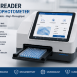
A plate reader spectrophotometer is one of the most useful instruments in modern laboratories. It helps scientists measure light absorbance, transmittance, or emission from samples placed in small wells on a plastic plate. This instrument saves time, reduces sample volume, and allows many tests to run together instead of one at a time.
Students, laboratory technologists, researchers, and healthcare workers use this device daily. It supports diagnostic testing, drug research, protein studies, and quality control in hospitals and pharmaceutical companies. If you are new to laboratory science, understanding this device will help you follow many other topics in biochemistry, microbiology, and immunology.
Definition
A plate reader spectrophotometer is a laboratory instrument that measures how much light passes through, is absorbed by, or is emitted from liquid samples held in the wells of a microplate. A microplate, also called a microtiter plate, is a flat plate with multiple small wells, commonly arranged in 96, 384, or even 1536 positions.
This instrument is also known by other names, including:
- Microplate reader
- Microplate spectrophotometer
- ELISA reader (when used mainly for enzyme-linked immunosorbent assays)
- Absorbance plate reader
Whatever name is used, the core function stays the same. The device shines a beam of light through each well and measures how the sample changes that light. From this measurement, scientists calculate the concentration, activity, or presence of a substance.
History
The story of the plate reader spectrophotometer began with the development of the microplate itself. In the 1950s, a Hungarian scientist named Gyula Takátsy designed one of the earliest multi-well plates to make virus testing faster and more efficient.
Over the following decades, laboratories realized that reading many small samples by hand was slow and prone to mistakes. This need pushed engineers to build automated reading instruments. By the 1970s and 1980s, the first dedicated microplate readers appeared, mainly to support the growing use of ELISA testing in immunology and virology.
As technology advanced, plate reader spectrophotometer models became faster, more sensitive, and capable of reading multiple wavelengths. Today’s instruments use advanced optics, computer software, and robotics to process hundreds of samples within minutes, something that was unimaginable with early manual methods.
Classification
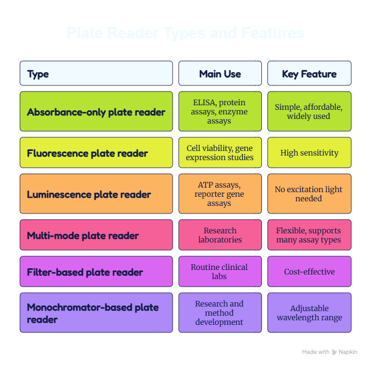
Plate readers can be classified in different ways based on their detection method and design.
Classification by Detection Method
- Absorbance plate readers – Measure how much light is absorbed by the sample. Most common type for ELISA and protein assays.
- Fluorescence plate readers – Detect light emitted by fluorescent molecules after excitation.
- Luminescence plate readers – Measure light produced by chemical or biological reactions without needing external excitation light.
- Multi-mode plate readers – Combine two or more detection methods (absorbance, fluorescence, and luminescence) in a single device.
Classification by Optical System
- Filter-based readers – Use physical filters to select specific wavelengths.
- Monochromator-based readers – Use a diffraction grating to select wavelengths, offering more flexibility.
Types of Plate Reader Spectrophotometer
Below is a simple comparison table of common types.
Components and Parts
A typical plate reader spectrophotometer contains several important parts working together.
- Light source: Usually a xenon flash lamp or tungsten-halogen lamp that produces light across a range of wavelengths.
- Wavelength selector: A filter or monochromator that isolates the specific wavelength needed for the test.
- Sample chamber (plate carrier): Holds the microplate steady during reading.
- Detector: A photodiode or photomultiplier tube (PMT) that measures the light after it passes through the sample.
- Optical fibers or mirrors: Direct light from the source to the sample and then to the detector.
- Microprocessor and software: Converts raw signals into readable data, such as absorbance values or concentration units.
- Display or connected computer: Shows the results in numeric or graphical form.
Shaking mechanism: Many modern readers include a built-in shaker to mix samples before reading.
Principle
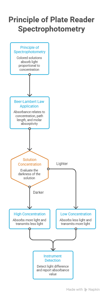
The plate reader spectrophotometer works on the basic principle of spectrophotometry. This principle states that a colored solution absorbs light in proportion to its concentration. This relationship is described by the Beer-Lambert Law, which says that absorbance is directly related to the concentration of the substance, the path length of light through the sample, and a constant called the molar absorptivity.
In simple words, a darker or more concentrated solution absorbs more light and lets less light pass through. The instrument detects this difference and reports it as an absorbance value.
Working Principle and Mechanism
Here is how the plate reader spectrophotometer works step by step:
- The light source produces a beam containing a range of wavelengths.
- The wavelength selector isolates the specific wavelength needed for the assay.
- This selected light beam passes through the sample in each well of the microplate.
- When the light beam passes through the sample, some of its energy is absorbed by the sample molecules, whereas the rest is transmitted.
- The detector measures the intensity of light that passes through.
- The microprocessor compares this value to a reference (blank) reading and calculates absorbance.
- Results appear on the screen as numeric values, and software can convert these into concentration using a standard curve.
This entire mechanism happens automatically and quickly, often reading a full 96-well plate within seconds.
Procedure
A general procedure for using a plate reader spectrophotometer includes the following steps:
- Preparation: Prepare samples, standards, and reagents according to the assay protocol.
- Plate loading: Add samples into the correct wells of the microplate using proper pipetting technique.
- Incubation: Allow the reaction to develop for the recommended time and temperature.
- Blanking: Set a blank well containing only the diluent or reagent without the sample to zero the instrument.
- Wavelength selection: Choose the correct wavelength based on the assay kit instructions.
- Plate insertion: Place the microplate carefully into the plate carrier.
- Reading: Start the reading process through the connected software.
- Data analysis: Review absorbance values, generate a standard curve if needed, and calculate final results.
- Reporting: Record and validate results before releasing them for clinical or research use.
Applications
The plate reader spectrophotometer supports many fields of science and medicine.
- Clinical diagnostics: Detecting infections such as HIV, hepatitis, and dengue through ELISA testing.
- Immunology research: Measuring antibody levels and immune response.
- Microbiology: Monitoring bacterial growth curves.
- Biochemistry: Measuring enzyme activity and protein concentration.
- Pharmaceutical industry: Drug screening and quality testing.
- Molecular biology: DNA and RNA quantification when combined with specific reagents.
- Toxicology: Studying cell viability and toxic effects of substances.
Food and environmental testing: Detecting contaminants or nutrient levels.
Advantages
- Allows testing of many samples at once, saving time.
- Requires very small sample volumes.
- Provides objective, numeric results instead of visual judgment.
- Reduces human error compared to manual reading methods.
- Supports multiple assay types with multi-mode instruments.
- Easily integrates with laboratory information systems (LIS).
Disadvantages
- Initial purchase cost can be high, especially for multi-mode readers.
- Requires regular calibration and maintenance.
- Sensitive to dust, dirt, or scratches on the microplate.
- Errors in pipetting or plate handling can affect all wells.
- Some models need specific consumables, such as branded microplates.
Clinical Significance
The plate reader spectrophotometer plays a major role in clinical laboratories, especially in serology and immunology departments. It is central to ELISA testing, a widely used method for detecting antibodies or antigens related to infections such as HIV, hepatitis B and C, syphilis, and dengue fever.
Because this instrument gives numeric, reproducible results, it supports accurate diagnosis and monitoring of disease. Hospitals also use it for hormone testing, tumor marker analysis, and allergy testing. In pharmaceutical quality control, it helps confirm that drug batches meet safety standards before reaching patients.
Interpretation of Results
Interpreting results from a plate reader spectrophotometer depends on the assay type, but general rules apply.
- Absorbance values are usually compared against a cut-off value provided by the test kit.
- A sample with absorbance higher than the cut-off is often considered positive (for many ELISA assays).
- A sample with absorbance lower than the cut-off is often considered negative.
- Some tests require calculating a ratio (sample absorbance divided by cut-off) for a clearer interpretation.
- Standard curves, built using known concentrations, help convert absorbance into actual concentration values for quantitative assays.
Result Type | Absorbance Compared to Cut-off | Common Interpretation |
Positive | Higher | Antigen or antibody detected |
Negative | Lower | No significant detection |
Equivocal/Borderline | Close to cut-off | Retesting recommended |
Frequently Observed Errors
Laboratory professionals commonly encounter these errors while using a plate reader spectrophotometer:
- Air bubbles in wells causing false high or low readings.
- Incorrect blanking, leading to inaccurate absorbance values.
- Dirty or scratched microplates scattering light incorrectly.
- Wavelength mismatch, using the wrong filter for the assay.
- Uneven pipetting volumes across wells.
- Plate misalignment inside the carrier.
- Lamp aging, reducing light intensity over time.
- Condensation on the plate lid affecting readings.
Troubleshooting
Problem | Possible Cause | Suggested Solution |
No reading or blank screen | Lamp failure or power issue | Check lamp status and power connections |
Inconsistent results between wells | Air bubbles or pipetting error | Repeat pipetting carefully, avoid bubbles |
High background absorbance | Dirty microplate or reagent contamination | Use clean plates and fresh reagents |
Error message on software | Wrong plate type selected | Match plate type in software settings |
Sudden drop in signal | Lamp aging | Replace lamp as per manufacturer schedule |
Care and Maintenance
Proper care extends the life and accuracy of a plate reader spectrophotometer.
- Clean the plate carrier regularly with a soft, lint-free cloth.
- Avoid touching optical components directly.
- Keep the instrument in a dust-free, vibration-free area.
- Follow the manufacturer’s schedule for lamp replacement.
- Perform regular calibration using certified reference standards.
- Store the instrument cover closed when not in use to prevent dust accumulation.
Quality Assurance (IQC and EQA)
Quality assurance is essential for ensuring that a plate reader spectrophotometer consistently produces accurate, reliable, and reproducible results throughout its service life.
Internal Quality Control (IQC)
- Running known positive and negative controls with every test batch.
- Checking absorbance values of controls against expected ranges.
- Documenting daily calibration checks.
- Recording lamp performance and wavelength accuracy periodically.
External Quality Assessment (EQA)
- Participating in national or international proficiency testing programs.
- Comparing laboratory results with other laboratories using the same test method.
- Identifying and correcting any systematic errors detected through EQA reports.
Following both IQC and EQA practices supports trustworthy, evidence-based laboratory testing, which is essential for patient safety.
Safety Precautions
- Always wear a lab coat and gloves while handling biological samples.
- Dispose of used micro plates as biohazard waste following local guidelines.
- Avoid looking directly at the light source during maintenance.
- Switch off and unplug the instrument before internal cleaning.
- Follow the manufacturer’s manual for electrical safety.
Handle reagents according to their material safety data sheets (MSDS).
Key Takeaways
- A plate reader spectrophotometer measures light absorbance, fluorescence, or luminescence from samples in microplate wells.
- It works based on the Beer-Lambert Law, linking light absorbance to sample concentration.
- Common types include absorbance-only, fluorescence, luminescence, and multi-mode readers.
- This instrument is essential in clinical diagnostics, especially for ELISA-based infection testing.
- Regular calibration, cleaning, and quality control keep results accurate and trustworthy.
- Understanding proper procedure and troubleshooting reduces common laboratory errors.
Conclusion
The plate reader spectrophotometer stands as a cornerstone instrument in modern laboratory science. From its early roots in microplate design to today’s advanced multi-mode systems, this device has transformed how scientists and clinicians test large numbers of samples quickly and accurately.
By understanding its principle, working mechanism, procedure, and quality control practices, students and professionals can use this instrument confidently and interpret results correctly. Whether used for diagnosing infections, studying enzyme activity, or supporting pharmaceutical research, the plate reader spectrophotometer continues to play a trustworthy role in advancing health and science.
As laboratory technology keeps evolving, mastering the fundamentals of this instrument prepares learners and professionals for more advanced tools and techniques in the future.
Frequently Asked Questions (FAQs)
What is a plate reader spectrophotometer used for?
It is used to measure light absorbance, fluorescence, or luminescence in samples placed in microplate wells, supporting tests like ELISA, enzyme assays, and protein quantification.
How does a plate reader spectrophotometer work?
It shines a selected wavelength of light through each sample well and measures how much light is absorbed, then converts this into readable data using the Beer-Lambert Law.
What is the difference between a plate reader and a regular spectrophotometer ?
A regular spectrophotometer reads one sample in a cuvette at a time, while a plate reader spectrophotometer reads many samples at once in the wells of a microplate.
Can a plate reader spectrophotometer detect DNA or RNA?
Some plate readers, especially UV-compatible models, can estimate nucleic acid concentration, though dedicated instruments like nanodrop spectrophotometers are more common for this purpose.
Why is blanking important before reading a microplate?
Blanking removes background signal from the diluent or reagent alone, ensuring the reported absorbance reflects only the sample of interest.
What is the normal wavelength used for ELISA testing?
Most ELISA kits use wavelengths between 405 nm and 450 nm, though the exact value depends on the specific test kit instructions.
How often should a plate reader spectrophotometer be calibrated?
Calibration frequency depends on the manufacturer’s guidelines and laboratory accreditation standards, but many labs perform checks daily and full calibration periodically, such as monthly or quarterly.
What causes inconsistent readings in a plate reader spectrophotometer?
Common causes include air bubbles, pipetting errors, dirty plates, incorrect wavelength selection, and lamp aging.
Is a plate reader spectrophotometer only used in hospitals?
No, it is also widely used in research laboratories, pharmaceutical companies, food testing facilities, and academic institutions.
Disclaimer
The information and materials provided on MeroLab are intended solely for educational, informational, professional learning, and public health awareness purposes. The website may contain content related to medical laboratory science, laboratory techniques, diagnostic tests, medical devices and instruments, reagents, diseases and health conditions, laboratory procedures, quality management, research, exam preparation, MCQs, and other healthcare-related topics.