
Introduction
A fluorescence microscope is one of the most powerful tools used in modern biology, medicine, and research laboratories. Unlike ordinary microscopes, it allows scientists to see specific structures inside cells with remarkable clarity and detail.
This instrument has transformed the way researchers study cells, proteins, and microorganisms. In this article, we will explore the fluorescence microscope in depth — covering its history, classification, types, parts, working principle, uses, and proper care and maintenance.
By the end of this guide, you will have a complete and practical understanding of why the fluorescence microscope remains an essential instrument in today’s scientific world.
What Is a Fluorescence Microscope?
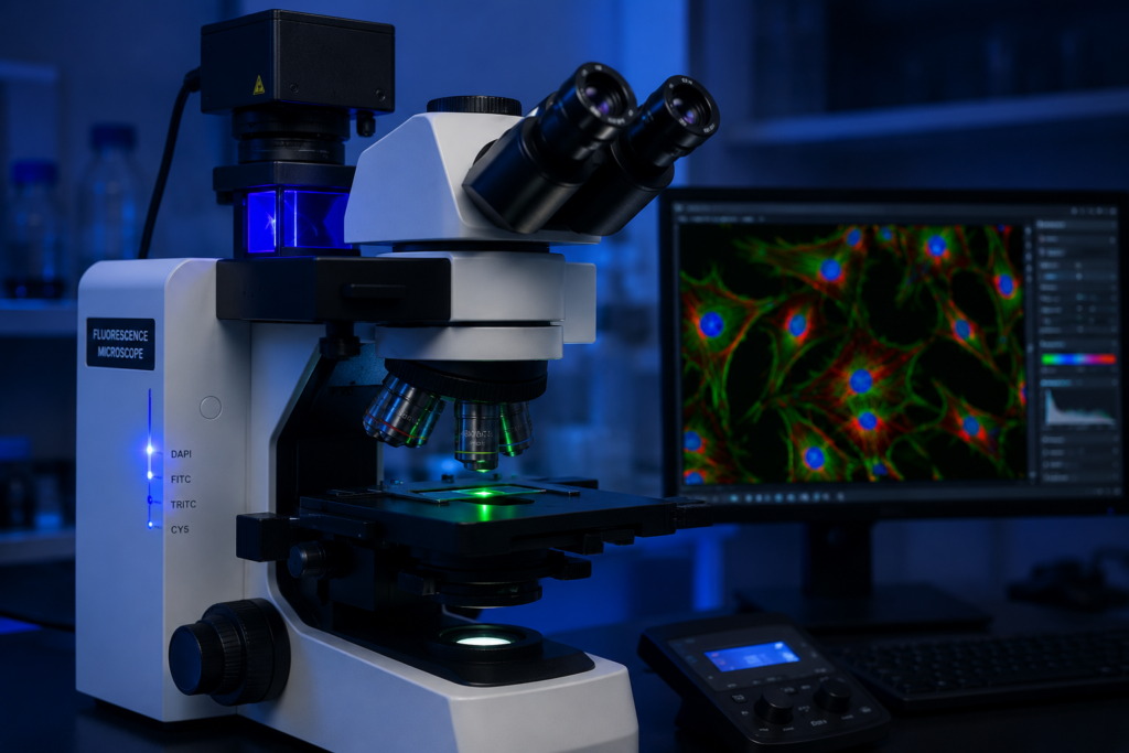
A fluorescence microscope is an advanced optical microscope that uses fluorescence instead of, or in addition to, simple reflection or absorption of light to study the properties of a specimen. It uses a high-intensity light source to excite fluorescent molecules, called fluorophores, within the sample.
When these fluorophores absorb light of a specific wavelength, they emit light of a longer wavelength, creating a glowing image against a dark background. This unique feature makes the fluorescence microscope extremely useful for identifying specific cell structures that would otherwise remain invisible under a normal microscope.
History and Background of Fluorescence Microscope
The concept of fluorescence microscopy dates back to the early 20th century. In 1911, German scientist Oskar Heimstädt built the first fluorescence microscope to study autofluorescence in biological specimens.
Over the following decades, researchers improved the design by introducing better light filters and more powerful light sources. The development of fluorescent dyes and antibodies in the mid-20th century further expanded the capabilities of the fluorescence microscope, allowing scientists to label specific proteins and structures within cells.
Today, the fluorescence microscope has evolved into a highly sophisticated tool, forming the foundation for advanced techniques such as confocal and super-resolution microscopy.
Classification of Fluorescence Microscope
The fluorescence microscope can be classified based on illumination method, design structure, and imaging technique.
- Based on Illumination Method
- Transmitted Fluorescence Microscope – Light passes through the specimen from below.
- Epi-Fluorescence Microscope – Light is directed onto the specimen from above through the objective lens; this is the most common modern design.
- Based on Light Source
- Mercury Vapor Lamp Microscope – Uses mercury lamps for high-intensity illumination.
- Xenon Lamp Microscope – Provides a broader and more stable light spectrum.
- LED-Based Microscope – Uses light-emitting diodes for longer lifespan and energy efficiency.
- Based on Imaging Technique
- Widefield Fluorescence Microscope – Illuminates the entire specimen at once.
- Confocal Fluorescence Microscope – Uses point-by-point scanning for sharper, highresolution images.
Types of Fluorescence Microscope
There are several types of fluorescence microscope, each suited for different research and diagnostic needs.
Type Common Use
Epi-Fluorescence Microscope | General cell and tissue imaging |
Confocal Microscope | High-resolution 3D imaging |
Total Internal Reflection Fluorescence (TIRF) Microscope | Studying cell surface events |
Super-Resolution Microscope | Imaging structures beyond the diffraction limit |
Multiphoton Microscope | Deep tissue imaging with reduced photodamage |
Each type of fluorescence microscope has been developed to address specific limitations of traditional light microscopy, offering researchers greater precision and flexibility.
Parts of a Fluorescence Microscope
Understanding the parts of a fluorescence microscope is important for accurate operation and image interpretation.
Mechanical Parts
- Base – Provides stability and support to the entire microscope.
- Arm – Connects the base to the body and eyepiece.
- Stage – Platform where the specimen slide is placed.
- Focus Knobs – Coarse and fine adjustment knobs for sharp focusing.
- Optical and Illumination Parts
- Light Source – Mercury, xenon, or LED lamp that provides high-intensity excitation light.
- Excitation Filter – Selects the specific wavelength of light needed to excite the fluorophore.
- Dichroic Mirror – Reflects excitation light toward the specimen while allowing emitted light to pass through toward the detector.
- Objective Lens – Focuses light onto the specimen and collects emitted fluorescence.
- Emission Filter – Filters out unwanted wavelengths, allowing only the emitted fluorescent light to reach the eyepiece or camera.
- Eyepiece (Ocular Lens) – Magnifies the final image for viewing.
- Detector or Camera – Captures digital images for analysis and documentation.
Working Principle of Fluorescence Microscope
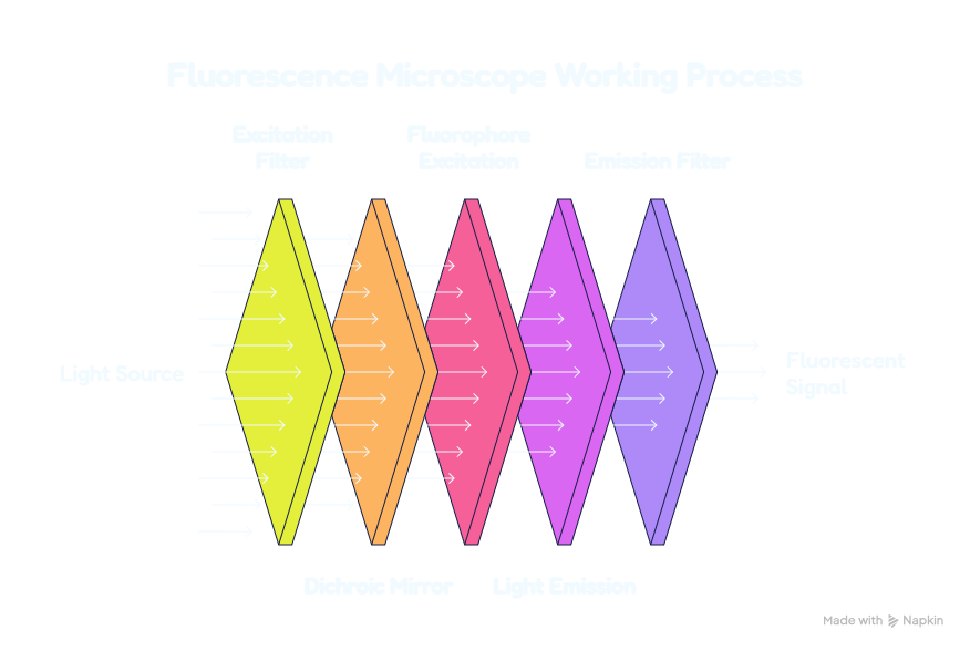
The working principle of a fluorescence microscope is based on the phenomenon of fluorescence, where certain molecules absorb light at one wavelength and emit light at a longer wavelength.
Step-by-Step Working Process
- Light from the source passes through the excitation filter, allowing only a specific wavelength to pass.
- This excitation light is reflected by the dichroic mirror toward the specimen.
- Fluorophores in the specimen absorb the excitation light and become energetically excited.
- As the fluorophores return to their normal state, they emit light of a longer wavelength.
- This emitted light passes back through the objective lens and dichroic mirror.
- The emission filter blocks any remaining excitation light, allowing only the fluorescent signal to reach the eyepiece or detector.
This process creates a bright, glowing image of specific structures against a dark background, offering excellent contrast and specificity.
Uses of Fluorescence Microscope
The fluorescence microscope is widely used across various scientific and medical fields due to its high specificity and sensitivity.
In Research
Studying protein localization within cells
Observing gene expression using fluorescent markers
Investigating cell structure and function
In Healthcare and Diagnostics
Identifying specific pathogens using fluorescent antibodies
Diagnosing autoimmune diseases through immunofluorescence testing
Detecting cancer cells in tissue samples
In Microbiology
Studying bacterial and viral structures
Observing live-cell processes in real time
In Neuroscience
Mapping neural connections
Studying brain tissue at a cellular level
Importance of Fluorescence Microscope in Modern Science
The fluorescence microscope has become an indispensable tool in laboratories worldwide. Its ability to highlight specific molecules within complex biological samples has significantly advanced our understanding of cell biology, disease mechanisms, and drug development.
Without the fluorescence microscope, many breakthroughs in immunology, genetics, and cancer research would not have been possible. It continues to play a critical role in both clinical diagnostics and cutting-edge scientific discovery.
Advantages of Fluorescence Microscope
Offers high specificity by targeting particular molecules or structures
Provides excellent contrast against a dark background
Enables visualization of live cellular processes
Supports multiple labeling techniques for studying different components simultaneously
Forms the foundation for advanced imaging techniques like confocal microscopy
Limitations of Fluorescence Microscope
Photobleaching can cause fluorescent signals to fade over time
Requires specialized and often expensive equipment
Sample preparation can be more complex compared to standard microscopy
Excessive light exposure may damage live samples
Care and Maintenance of Fluorescence Microscope
Proper care and maintenance of a fluorescence microscope is essential to preserve image quality and extend the instrument’s working life.
Daily Care Tips
Always turn off the light source when not in use to extend lamp life.
Clean optical components gently using lens paper only.
Avoid exposing filters and mirrors to dust or moisture.
Cover the microscope after each use.
Maintenance Best Practices
Replace mercury or xenon lamps according to the manufacturer’s recommended schedule.
Regularly check and clean excitation and emission filters.
Store the microscope in a dark, temperature-controlled environment.
Handle the objective lenses carefully to avoid scratches.
Schedule periodic professional servicing for calibration and alignment.
Avoid prolonged exposure of samples to excitation light to reduce photobleaching.
Following these maintenance steps helps ensure consistent image quality and prolongs the lifespan of this valuable instrument.
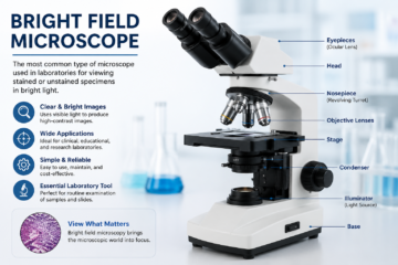
Difference Between Fluorescence Microscope and Bright Field Microscope
Feature | Fluorescence Microscope | Bright Field Microscope |
Light Source | High-intensity excitation light | Standard white light |
Background | Dark | Bright |
Specificity | High, targets specific molecules | Low, general observation |
Common Use | Advanced research and diagnostics | General education and basic diagnostics |
Cost | Higher | Lower |
This comparison shows why the fluorescence microscope is preferred for specialized research applications, while bright field microscopes remain suitable for general laboratory use.
Why Fluorescence Microscope Remains Essential Today
Despite the rise of newer imaging technologies, the fluorescence microscope continues to be a cornerstone of biological and medical research. Its unmatched ability to visualize specific molecules within complex samples makes it irreplaceable in many scientific fields.
From diagnosing diseases to exploring the fundamental mechanisms of life, the fluorescence microscope empowers researchers and clinicians to make discoveries that were once impossible.
For further reading on fluorescence microscopy techniques and applications, professionals can explore trusted academic resources such as PubMed and PMC, which offer peerreviewed research on microscopy advancements in clinical and laboratory settings.
Why Fluorescence Microscope Remains Essential Today
Despite the rise of newer imaging technologies, the fluorescence microscope continues to be a cornerstone of biological and medical research. Its unmatched ability to visualize specific molecules within complex samples makes it irreplaceable in many scientific fields.
From diagnosing diseases to exploring the fundamental mechanisms of life, the fluorescence microscope empowers researchers and clinicians to make discoveries that were once impossible.
For further reading on fluorescence microscopy techniques and applications, professionals can explore trusted academic resources such as PubMed and PMC, which offer peerreviewed research on microscopy advancements in clinical and laboratory settings.
Conclusion
The fluorescence microscope stands as one of the most powerful and transformative tools in modern science. From its fascinating history to its detailed working principle, this instrument has reshaped how we study cells, tissues, and disease at a molecular level.
Understanding its classification, types, parts, uses, and proper care ensures that students, researchers, and healthcare professionals can make the most of this remarkable technology while extending its operational lifespan.
Frequently Asked Questions (FAQs)
What is a fluorescence microscope used for?
A fluorescence microscope is used to visualize specific molecules, proteins, and structures within cells and tissues using fluorescent markers.
What is the main working principle of a fluorescence microscope?
It works by exciting fluorophores in a specimen with a specific wavelength of light, causing them to emit light of a longer wavelength, which is then captured to form an image.
What are the main parts of a fluorescence microscope?
Key parts include the light source, excitation filter, dichroic mirror, objective lens, emission filter, and detector or camera.
How can I maintain a fluorescence microscope properly?
Turn off the light source when not in use, clean optical parts with lens paper, replace lamps as recommended, and schedule regular professional servicing.
What is the difference between a fluorescence microscope and a confocal microscope?
A fluorescence microscope illuminates the entire specimen at once, while a confocal microscope uses point-by-point scanning to produce sharper, high-resolution three-dimensional images.
Why is the fluorescence microscope important in medical research?
It allows researchers to study specific cell structures, diagnose diseases through immunofluorescence, and investigate molecular processes that are invisible under standard microscopes.