
Introduction
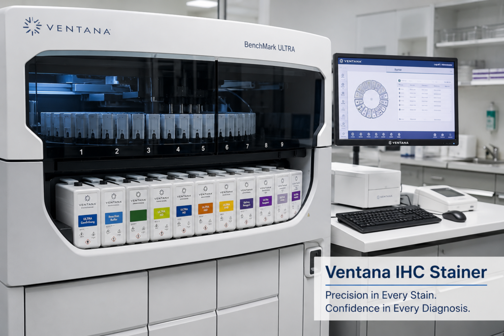
The Ventana IHC stainer is one of the most important instruments used in modern pathology laboratories today. This automated machine helps scientists and pathologists stain tissue samples so they can detect specific proteins inside cells. These proteins, called antigens, often reveal important clues about diseases such as cancer, infections, and autoimmune disorders.
Before automation, immunohistochemistry (IHC) staining was done by hand. This manual process was slow, inconsistent, and prone to human error. The introduction of the Ventana IHC stainer changed this completely. Made by Roche Diagnostics under the Ventana Medical Systems brand, this instrument brought speed, accuracy, and reliability to tissue staining.
Definition
The Ventana IHC stainer is an automated laboratory instrument designed to perform immunohistochemistry (IHC) and in situ hybridization (ISH) staining on tissue sections placed on glass microscope slides.
Immunohistochemistry (IHC) is a laboratory technique that uses antibodies to detect specific antigens (target proteins) in tissue samples. When an antibody binds to its matching antigen, a colored marker appears under the microscope, showing exactly where that protein is located in the tissue.
The Ventana IHC stainer automates every step of this process, including:
- Baking and dewaxing tissue sections
- Antigen retrieval (unmasking hidden proteins)
- Applying primary and secondary antibodies
- Adding detection chemicals
- Counterstaining the tissue
This automation reduces human error and produces consistent, high-quality results every time.
History
Understanding the history of the Ventana IHC stainer helps us appreciate how far pathology testing has come.
- 1985: Ventana Medical Systems was founded in Arizona, USA, with a goal of automating tissue diagnostics.
- 1991: Ventana launched one of the first automated IHC staining systems, moving the field away from fully manual staining.
- 2000s: Ventana introduced the BenchMark series, including BenchMark XT and BenchMark Ultra, which combined IHC and ISH staining capabilities in one platform.
- 2008: Roche Diagnostics acquired Ventana Medical Systems, expanding its global reach and integrating the technology into Roche’s broader diagnostics portfolio.
- Present day: The Ventana BenchMark Ultra and BenchMark Ultra PLUS remain widely used in hospitals and reference laboratories worldwide, supporting both research and clinical diagnostic testing.
This steady evolution reflects the growing demand for precise, reproducible, and efficient tissue-based diagnostics, especially in cancer care.
Classification
Ventana IHC stainers can be classified based on different factors:
1. Based on Automation Level
- Semi-automated systems – Require some manual steps before or after staining.
- Fully automated systems – Perform the entire staining process, including baking, dewaxing, antigen retrieval, and staining, without manual intervention.
2. Based on Staining Capability
- IHC-only stainers – Perform immunohistochemistry staining only.
- Dual IHC/ISH stainers – Perform both immunohistochemistry and in situ hybridization staining, such as the BenchMark Ultra.
3. Based on Throughput
- Low-throughput instruments – Suitable for small laboratories with fewer daily samples.
- High-throughput instruments – Designed for large reference laboratories processing hundreds of slides daily.
Types of Ventana IHC Stainers
Several models have been developed under the Ventana brand. Below is a comparison table of the most common types.
Model Name | Key Feature | Best Suited For |
Ventana BenchMark XT | Fully automated IHC and ISH staining | Mid-to-large pathology labs |
Ventana BenchMark Ultra | Advanced automation with barcode tracking | High-volume reference laboratories |
Ventana BenchMark Ultra PLUS | Enhanced software and workflow integration | Large hospital networks |
Ventana BenchMark GX | Compact design for smaller labs | Small to mid-size laboratories |
Ventana Discovery Ultra | Research-focused staining platform | Research and translational studies |
Each model shares the same core staining principle but differs in slide capacity, software features, and integration with laboratory information systems (LIS).
Components / Parts of a Ventana IHC Stainer
A Ventana IHC stainer is made up of several important parts that work together to complete the staining process.
- Slide Tray/Carousel – Holds glass slides in position during processing.
- Reagent Dispensing System – Automatically applies antibodies, detection kits, and buffers onto each slide.
- Heating Module – Controls temperature for tissue baking and antigen retrieval.
- Liquid Coverslip (LCS) System – Applies a thin oil layer over reagents to prevent evaporation during incubation.
- Barcode Scanner – Reads slide barcodes to match each sample with its correct staining protocol.
- Touchscreen Interface – Allows technologists to program, monitor, and control staining runs.
- Waste Management System – Safely collects used reagents and buffers.
- Software System (e.g., VANTAGE or NexES) – Manages protocols, tracks results, and connects with laboratory databases.
Principle
The Ventana IHC stainer works on the basic principle of antigen-antibody interaction.
In simple terms:
- Every cell contains specific proteins (antigens).
- A matching antibody is designed to recognize and bind only to that specific antigen.
- Once bound, a detection system (usually enzyme-linked) produces a visible color change.
- This colored signal shows exactly where the target protein exists in the tissue.
This principle allows pathologists to identify disease markers, such as hormone receptors in breast cancer or specific viral proteins in infected tissue.
Working Principle and Mechanism
The working mechanism of the Ventana IHC stainer follows a well-organized sequence of automated steps.
Step 1: Deparaffinization
Tissue samples are usually embedded in paraffin wax before sectioning. The stainer heats the slides to melt and remove this wax, exposing the tissue underneath.
Step 2: Antigen Retrieval
Formalin fixation (a chemical process used to preserve tissue) can hide or “mask” antigens. The instrument uses heat-induced or enzyme-based antigen retrieval to unmask these proteins so antibodies can bind properly.
Step 3: Primary Antibody Application
A primary antibody, specific to the target antigen, is automatically dispensed onto the tissue section.
Step 4: Secondary Antibody and Detection
A secondary antibody, linked to an enzyme (commonly horseradish peroxidase), binds to the primary antibody. This enzyme reacts with a chromogen (a colorless substance that turns colored), producing a visible brown or red stain, depending on the detection kit used.
Step 5: Counterstaining
A counterstain, usually hematoxylin, is applied to color the cell nuclei blue. This provides contrast, making it easier to see tissue structure under the microscope.
Step 6: Liquid Coverslip Application
A thin oil-based liquid coverslip is applied over the reagents to prevent evaporation and maintain consistent reaction conditions.
Step 7: Slide Unloading
Once staining is complete, slides are removed, cleaned of excess reagent, and prepared for microscopic examination by a pathologist.
Procedure: Step-by-Step Workflow
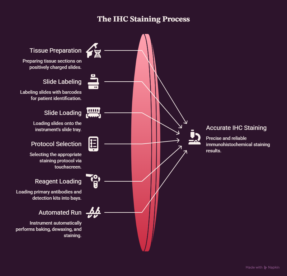
Here is a simplified overview of how laboratory staff typically operate a Ventana IHC stainer:
- Prepare tissue sections on positively charged glass slides.
- Label slides with barcodes matching patient and test information.
- Load slides onto the instrument’s slide tray.
- Select the staining protocol through the touchscreen or barcode scan.
- Load reagents, including primary antibodies and detection kits, into designated reagent bays.
- Start the automated run. The instrument performs baking, dewaxing, antigen retrieval, staining, and counterstaining automatically.
- Monitor the run using the software interface for any error alerts.
- Unload slides once the cycle finishes.
- Perform manual dehydration and coverslipping (in most models) before microscopic review.
- Review slides under a microscope for interpretation by a qualified pathologist.
Applications
The Ventana IHC stainer is used across many areas of medicine and research.
- Cancer diagnosis – Identifying tumor markers such as HER2, ER, PR, and Ki-67 in breast cancer.
- Infectious disease detection – Detecting viral or bacterial antigens in infected tissue.
- Autoimmune disease evaluation – Identifying immune complex deposits in tissue biopsies.
- Transplant pathology – Assessing organ rejection markers.
- Research studies – Supporting translational and pharmaceutical research on new biomarkers.
- Companion diagnostics – Helping determine which patients are eligible for targeted cancer therapies.
Advantages
The Ventana IHC stainer offers several benefits over manual staining methods.
- Consistency – Reduces variation between staining runs.
- Time-saving – Completes staining in hours instead of an entire day.
- Reduced human error – Minimizes mistakes from manual pipetting and timing.
- Standardization – Ensures reproducible results across different technicians and shifts.
- Traceability – Barcode systems track each slide from start to finish.
- High throughput – Processes multiple slides simultaneously, ideal for busy laboratories.
- Improved safety – Reduces direct handling of chemicals and biological samples.
Disadvantages
Despite its many strengths, the Ventana IHC stainer also has some limitations.
- High cost – The initial purchase price and maintenance contracts can be expensive.
- Reagent dependency – Often requires proprietary Ventana-branded reagents, increasing running costs.
- Technical complexity – Requires trained personnel for operation and troubleshooting.
- Downtime risk – Mechanical or software errors can delay testing.
- Space requirement – The instrument requires dedicated bench or floor space in the lab.
Clinical Significance
The clinical importance of the Ventana IHC stainer cannot be overstated. Many life-changing medical decisions depend on accurate IHC results generated by this instrument.
- Breast cancer treatment planning relies heavily on HER2, ER, and PR status determined through IHC staining.
- Lymphoma classification depends on identifying specific cell surface markers.
- Infection diagnosis, such as detecting Helicobacter pylori in stomach biopsies, uses IHC-based confirmation.
- Personalized medicine increasingly depends on biomarker testing to match patients with the most effective targeted therapies.
Because these results directly influence treatment decisions, the accuracy and reliability provided by automated systems like the Ventana IHC stainer are critical to patient safety.
Interpretation of Results
Interpreting IHC results requires careful evaluation by a trained pathologist. Key factors include:
- Staining intensity – Weak, moderate, or strong staining reactions.
- Staining pattern – Nuclear, cytoplasmic, or membranous localization of the stain.
- Percentage of positive cells – The proportion of stained cells within the tissue sample.
- Comparison with controls – Positive and negative control slides help confirm that staining worked correctly.
Result Type | Description |
Positive Control | Confirms the staining reagents and process worked correctly |
Negative Control | Confirms there is no non-specific background staining |
True Positive | Target antigen is present, and staining appears as expected |
False Negative | Antigen is present, but staining fails due to technical error |
False Positive | Staining appears without true antigen presence, often due to background noise |
Frequently Observed Errors
Even with automation, errors can occur. Common issues include:
- Weak or absent staining – Often caused by expired reagents or insufficient antigen retrieval.
- High background staining – May result from inadequate washing steps or antibody concentration issues.
- Uneven staining across the slide – Can occur due to improper reagent dispensing or air bubbles.
- Barcode scanning errors – Caused by damaged or poorly printed labels.
- Reagent evaporation – Happens if the liquid coverslip system malfunctions.
Troubleshooting
Laboratories can resolve many common issues with these troubleshooting tips:
- Check reagent expiry dates regularly before starting a run.
- Inspect barcode labels for clarity and correct placement.
- Verify antigen retrieval settings match the manufacturer’s recommended protocol for each antibody.
- Calibrate the instrument according to the scheduled maintenance plan.
- Run control slides with every batch to confirm staining accuracy.
- Contact technical support promptly if software errors persist.
Care and Maintenance
Proper maintenance keeps the Ventana IHC stainer running efficiently and extends its service life.
- Daily maintenance – Clean dispensing tips and check reagent levels.
- Weekly maintenance – Inspect tubing systems for blockages or leaks.
- Monthly maintenance – Perform software updates and calibration checks.
- Annual maintenance – Schedule professional servicing by certified technicians.
- Environmental control – Keep the instrument in a stable, dust-free, temperature-controlled room.
Quality Assurance (IQC and EQA)
Quality assurance is essential to ensure the Ventana IHC stainer produces reliable and clinically trustworthy results.
Internal Quality Control (IQC)
- Running known positive and negative control tissues with every staining batch.
- Documenting reagent lot numbers and expiry dates.
- Recording daily instrument performance checks.
External Quality Assessment (EQA)
- Participating in national or international proficiency testing programs.
- Comparing laboratory results with peer laboratories to identify inconsistencies.
- Following accreditation standards, such as those set by CLSI (Clinical and Laboratory Standards Institute) and CAP (College of American Pathologists).
Consistent IQC and EQA practices help laboratories maintain accreditation and, most importantly, ensure patient safety.
Safety Precautions
Working with the Ventana IHC stainer involves handling chemicals and biological samples, so safety precautions are essential.
- Always wear appropriate personal protective equipment (PPE), such as gloves, a laboratory coat, and safety goggles or other eye protection.
- Handle reagents in a well-ventilated area, as some chemicals can be irritating.
- Dispose of biological waste according to local biosafety regulations.
- Avoid direct skin contact with staining reagents and chromogens.
- Follow manufacturer guidelines for instrument operation and emergency shutdown procedures.
- Report spills immediately and follow standard laboratory spill protocols.
Key Points
- The Ventana IHC stainer automates immunohistochemistry staining, improving speed, accuracy, and consistency.
- It works on the principle of antigen-antibody binding, followed by chemical detection.
- Common models include the BenchMark XT, BenchMark Ultra, and BenchMark Ultra PLUS.
- The instrument plays a critical role in cancer diagnosis, infection detection, and personalized medicine.
- Regular maintenance, quality control, and staff training are essential for reliable results.
- Despite higher costs, its benefits in standardization and diagnostic accuracy make it a valuable investment for pathology laboratories.
Conclusion
The Ventana IHC stainer has transformed the field of anatomic pathology by automating a process that was once slow, manual, and inconsistent. From its origins with Ventana Medical Systems to its current role under Roche Diagnostics, this instrument has become a trusted tool in laboratories worldwide.
By automating steps such as deparaffinization, antigen retrieval, antibody application, and counterstaining, the Ventana IHC stainer ensures that pathologists receive consistent, high-quality slides for accurate diagnosis. Its role in detecting cancer biomarkers, infectious agents, and other disease indicators makes it a cornerstone of modern diagnostic pathology.
While the instrument does require significant investment and proper maintenance, the benefits in accuracy, efficiency, and patient safety make it an essential part of today’s healthcare diagnostic landscape. As personalized medicine continues to grow, tools like the Ventana IHC stainer will remain vital in guiding effective, targeted patient treatment.
Frequently Asked Questions (FAQs)
What is a Ventana IHC stainer used for?
It is used to automatically stain tissue samples so pathologists can detect specific proteins (antigens) linked to diseases such as cancer and infections.
Who manufactures the Ventana IHC stainer?
It is manufactured by Ventana Medical Systems, a company that became part of Roche Diagnostics in 2008.
How long does an IHC staining run take on this instrument?
Staining runs typically take a few hours, though exact timing depends on the specific protocol and antibody used.
What is the difference between IHC and ISH staining?
IHC detects proteins using antibodies, while in situ hybridization (ISH) detects specific DNA or RNA sequences using labeled probes. Many Ventana instruments, like the BenchMark Ultra, can perform both.
Why is antigen retrieval necessary before staining?
Formalin fixation can hide antigens by cross-linking proteins. Antigen retrieval unmasks these proteins so antibodies can bind effectively during staining.
Can small laboratories afford a Ventana IHC stainer?
Smaller laboratories may consider compact models like the BenchMark GX, which offer automation at a lower throughput and cost compared to larger systems.
What controls are used to verify accurate staining?
Positive and negative control tissues are run alongside patient samples to confirm that the staining process worked correctly.
Is special training required to operate this instrument?
Yes, laboratory personnel typically undergo manufacturer-provided training to operate the instrument safely and interpret results correctly.
How does automation improve diagnostic accuracy?
Automation reduces variability caused by manual handling, ensuring more consistent reagent application, timing, and temperature control across every slide.