
Introduction:
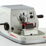
A Leica microtome is one of the most trusted and widely used instruments in histopathology and research laboratories around the world. It is designed to cut extremely thin sections of tissue for microscopic examination, forming the backbone of accurate histological diagnosis.
Known for its precision engineering and reliability, Leica has become a leading name in microtome manufacturing. In this article, we will explore the Leica microtome in depth—covering its history, classification, types, parts, working principle, uses, and proper care and maintenance.
By the end of this guide, you will have a complete and practical understanding of why the Leica microtome remains a preferred choice among pathologists, lab technicians, and researchers.
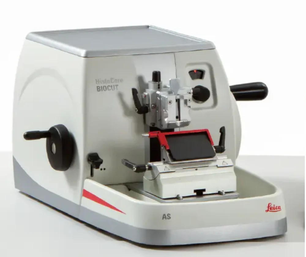
What Is a Leica Microtome?
A Leica microtome is a precision instrument manufactured by Leica Biosystems, used to cut thin, uniform slices of tissue samples, typically ranging from 1 to 10 microns in thickness. These thin sections are then mounted on slides and stained for examination under a microscope.
The Leica microtome is widely trusted in pathology laboratories because of its consistent cutting accuracy, durable build quality, and user-friendly design. It plays a critical role in preparing samples for cancer diagnosis, disease research, and routine histopathology testing.
History and Background of Leica Microtome
The history of the microtome dates back to the 18th century, when early instruments were hand-operated and limited in precision. Over time, engineers refined the design, introducing mechanical rotary systems for smoother and more accurate cutting.
Leica, a company with a long legacy in optical and scientific instruments, entered the field of microtomy by combining German engineering precision with advanced mechanical design. Today, the Leica microtome is recognized globally as a benchmark instrument in histology, offering improved accuracy, safety features, and ergonomic design compared to older manual models.
Modern Leica microtomes now include motorized and digital systems, reflecting decades of innovation in laboratory science.
Classification of Leica Microtome
The Leica microtome can be classified based on operation mode, cutting mechanism, and application.
- Based on Operation Mode
- Manual Microtome – Operated by hand using a rotating wheel.
- Semi-Automated Microtome – Combines manual control with motorized assistance.
- Fully Automated Microtome – Performs sectioning with minimal manual intervention, ideal for high-throughput laboratories.
- Based on Cutting Mechanism
- Rotary Microtome – Uses a rotating wheel to move the specimen against the blade.
- Sliding Microtome – The blade moves back and forth across a stationary specimen.
- Ultramicrotome – Used for extremely thin sections, often for electron microscopy.
- Based on Application
- Routine Histology Microtome – Used for standard tissue sectioning in diagnostic labs.
- Research-Grade Microtome – Designed for specialized studies requiring high precision.
Types of Leica Microtome
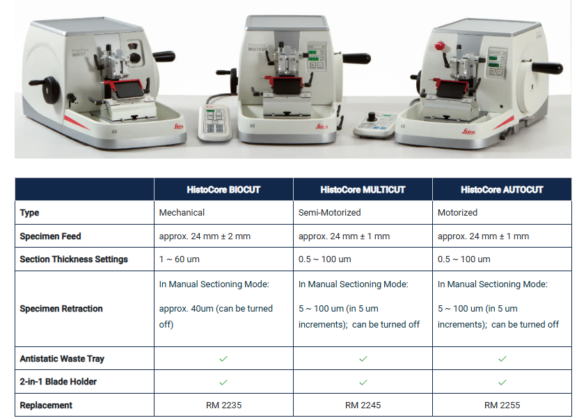
Leica offers several models of microtomes, each designed for specific laboratory needs.
Type Common Use
|
|
|
|
|
|
|
|
|
|
Each type of Leica microtome is engineered to balance precision, speed, and ease of use, depending on the laboratory’s workload and specific requirements.
Parts of a Leica Microtome
Understanding the parts of a Leica microtome is essential for proper operation and accurate sectioning.
Mechanical Parts
- Base – Provides stability and support to the entire instrument.
- Handwheel – Used to manually rotate and advance the specimen for cutting.
- Specimen Clamp – Holds the tissue block securely in place.
- Specimen Orientation System – Allows precise alignment of the tissue block.
- Section Thickness Control Dial – Adjusts the thickness of each tissue section, typically between 1 and 60 microns.
Cutting and Safety Parts
- Blade Holder – Secures the microtome blade firmly during cutting.
- Blade Guard – Protects the operator from accidental contact with the sharp blade.
- Waste Tray – Collects tissue waste and debris during sectioning.
- Retraction Mechanism – Moves the specimen away from the blade during the return stroke to prevent damage to sections.
Digital and Control Features (in Automated Models)
- Digital Display Panel – Shows section thickness and cutting speed settings.
- Foot Pedal Control – Allows hands-free operation in automated models.
Working Principle of Leica Microtome
The working principle of a Leica microtome is based on precise mechanical movement that advances a tissue specimen against a sharp blade in controlled, uniform increments.
Step-by-Step Working Process
- The tissue block, usually embedded in paraffin wax, is securely fixed in the specimen clamp.
- The section thickness is set using the control dial, depending on the required precision.
- As the handwheel is rotated (or the motorized system is activated), the specimen advances toward the blade.
- The blade slices through the tissue block, producing a thin, uniform section.
- The specimen retracts slightly on the return stroke to avoid damaging the blade or the tissue.
- This process is repeated to produce a continuous ribbon of tissue sections, which are then carefully transferred onto glass slides.
This controlled and repeatable process ensures that each tissue section is uniform in thickness, which is critical for accurate microscopic diagnosis.
Uses of Leica Microtome
The Leica microtome is used extensively across medical, research, and industrial fields.
In Histopathology
- Preparing tissue sections for cancer diagnosis
- Sectioning biopsy samples for disease identification
- Supporting routine diagnostic pathology workflows
In Research
- Studying tissue architecture in animal and plant samples
- Preparing samples for immunohistochemistry studies
- Supporting neuroscience and developmental biology research
In Forensic Science
- Sectioning tissue samples for forensic pathology investigations
In Industrial Applications
- Sectioning polymer and material samples for quality analysis
Importance of Leica Microtome in Modern Laboratories
The Leica microtome plays a vital role in ensuring accurate and reliable histological diagnosis. Since even minor inconsistencies in section thickness can affect staining quality and diagnostic accuracy, the precision offered by Leica microtomes is invaluable.
Pathologists depend on high-quality tissue sections to detect diseases such as cancer, infections, and organ abnormalities. Without a dependable microtome, the entire diagnostic process could be compromised, making this instrument a cornerstone of modern pathology laboratories.
Advantages of Leica Microtome
- Delivers highly precise and uniform tissue sections
- Durable build quality designed for long-term laboratory use
- Available in manual, semi-automated, and fully automated models
- Ergonomic design reduces operator fatigue during long sessions
- Trusted brand with strong global support and service network
Limitations of Leica Microtome
- Higher cost compared to generic microtome brands
- Requires proper training for optimal use, especially advanced models
- Regular maintenance is necessary to preserve cutting precision
- Blade handling requires strict safety precautions
Care and Maintenance of Leica Microtome
Proper care and maintenance of a Leica microtome ensures consistent performance and extends the instrument’s operational lifespan.
Daily Care Tips
- Clean the specimen clamp and surrounding area after each use to remove tissue debris.
- Always replace or sharpen blades regularly to maintain cutting precision.
- Keep the blade guard in place when the microtome is not actively in use.
- Wipe down the base and handwheel with a soft, dry cloth.
Maintenance Best Practices
- Lubricate moving mechanical parts as recommended by the manufacturer.
- Avoid exposing the microtome to excessive moisture or dust.
- Schedule periodic professional servicing to check calibration accuracy.
- Store the instrument in a stable, vibration-free environment.
- Train all operators on proper blade handling and safety protocols to prevent accidents.
Following these maintenance steps helps preserve cutting accuracy and ensures operator safety over the long-term use of the Leica microtome.
Difference Between Leica Microtome and Standard Microtome
Feature | Leica Microtome | Standard/Generic Microtome |
| High-precision German engineering | Varies by manufacturer |
| Manual, semi-automated, fully automated | Often limited to manual only |
| High section-to-section uniformity | May vary in accuracy |
| Strong global service network | Limited in some regions |
| Higher | Generally lower |
This comparison highlights why many diagnostic and research laboratories prefer the Leica microtome despite its higher initial investment, due to its long-term reliability and precision.
Why Leica Microtome Remains a Trusted Choice Today
Despite growing competition in laboratory instrumentation, the Leica microtome continues to be a preferred choice among pathologists and researchers worldwide. Its combination of precision, durability, and advanced automation options makes it suitable for both routine diagnostic work and specialized research applications.
As histopathology continues to advance, the Leica microtome remains an essential tool for producing consistent, high-quality tissue sections that support accurate diagnosis and scientific discovery.
For further reading on microtomy techniques and histopathology advancements, professionals can explore trusted academic resources such as PubMed and PMC, which offer peer-reviewed research on tissue processing and laboratory techniques.
Conclusion:
The Leica microtome stands as one of the most powerful and dependable instruments in modern histopathology and research laboratories. From its rich history to its precise working principle, this instrument continues to support accurate disease diagnosis and scientific advancement.
Understanding its classification, types, parts, uses, and proper care ensures that laboratory professionals and researchers can maximize both accuracy and equipment longevity.
Want to learn more about essential histology and laboratory instruments? Explore more expert guides on diagnostic tools and laboratory science to strengthen your practical knowledge.
Frequently Asked Questions (FAQs)
Q1: What is a Leica microtome used for?
It is used to cut extremely thin sections of tissue samples for microscopic examination in histopathology and research.
Q2: What is the main working principle of a Leica microtome?
It works by advancing a tissue specimen against a sharp blade in precise, controlled increments to produce uniform thin sections.
Q3: What are the main parts of a Leica microtome?
Key parts include the handwheel, specimen clamp, blade holder, section thickness control dial, and blade guard.
Q4: How can I maintain a Leica microtome properly?
Clean the instrument after each use, replace or sharpen blades regularly, lubricate moving parts, and schedule periodic professional servicing.
Q5: What is the difference between a manual and automated Leica microtome?
A manual microtome requires hand operation via a handwheel, while an automated Leica microtome uses a motorized system for faster, more consistent sectioning with less operator effort.
Q6: Why is the Leica microtome important in histopathology?
It ensures precise, uniform tissue sections, which are essential for accurate staining and reliable disease diagnosis under the microscope.