
Iron deficiency anemia is the most common nutritional deficiency worldwide — yet it is one of the most easily missed without proper laboratory investigation. Whether you are a laboratory technician processing samples, a clinician interpreting reports, a medical student studying hematology, or simply someone trying to understand your blood test results, this guide breaks down every key iron deficiency anemia lab test in clear, practical language.
What Is Iron Deficiency Anemia?
Iron deficiency anemia (IDA) develops when the body lacks enough iron to produce sufficient hemoglobin — the protein in red blood cells that carries oxygen to tissues. Without adequate hemoglobin, cells throughout the body receive less oxygen, causing fatigue, pallor, dizziness, and reduced work capacity.
IDA progresses through three well-defined stages, each detectable through laboratory testing:
Stage 1 — Iron Depletion
Body iron stores decline. Serum ferritin falls first. Hemoglobin remains normal at this point.
Stage 2 — Iron-Deficient Erythropoiesis
Less iron is available to developing red cells. Transferrin saturation drops. Red cell production is compromised.
Stage 3 — Iron Deficiency Anemia
Hemoglobin falls below normal. Microcytic, hypochromic red cells appear on the blood film.
Why Laboratory Tests Matter in IDA Diagnosis
Clinical signs alone — pale conjunctiva, koilonychia, fatigue — are suggestive but never confirmatory. Accurate diagnosis of iron deficiency anemia requires a panel of iron deficiency anemia labs that together paint a complete picture of iron metabolism. No single test is sufficient on its own; clinicians and laboratory professionals must interpret the full iron panel in context.
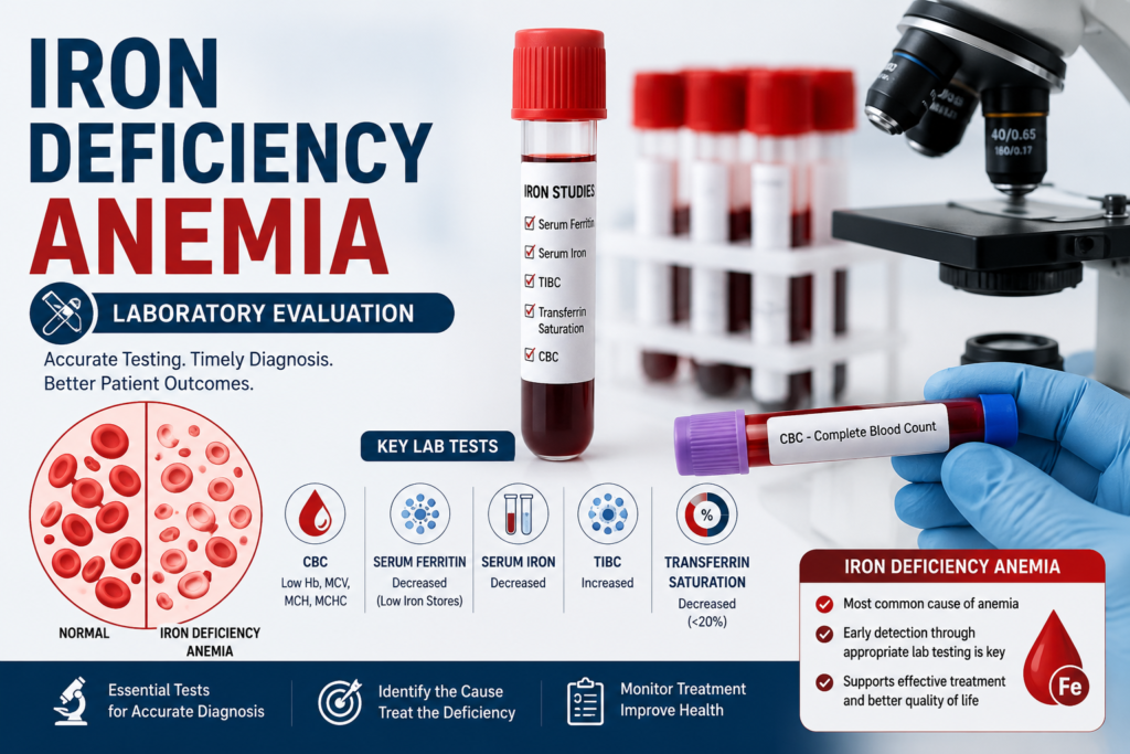
Core Iron Deficiency Anemia Lab Tests
The following tests form the backbone of any iron deficiency anemia laboratory workup. Each has a distinct working principle, reference range, and clinical meaning.
1. Complete Blood Count (CBC)
The CBC is the first and most widely ordered test in any suspected anemia. In iron deficiency anemia, the CBC typically reveals:
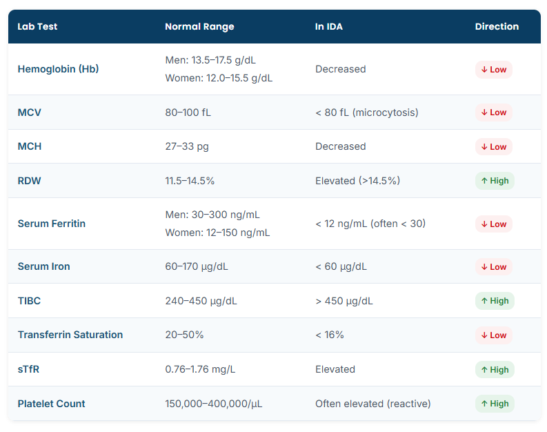
- Low hemoglobin (Hb): Below 13 g/dL in adult men; below 12 g/dL in adult women
- Low MCV (Mean Corpuscular Volume): Red cells are smaller than normal (microcytosis)
- Low MCH (Mean Corpuscular Hemoglobin): Red cells carry less hemoglobin (hypochromia)
- High RDW (Red Cell Distribution Width): Variability in red cell size increases (anisocytosis)
- Elevated platelet count: Reactive thrombocytosis is a common but often overlooked finding
Working Principle: The CBC is performed using an automated hematology analyzer (impedance or light-scatter technology). Red cells pass through an aperture or laser beam, generating electronic pulses that calculate cell volume, count, and hemoglobin concentration. The analyzer derives MCV, MCH, MCHC, and RDW from these measurements.
2. Peripheral Blood Film (PBS)
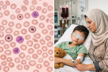
A peripheral blood smear provides a direct visual picture of red cell morphology. In iron deficiency anemia, a trained laboratory technologist observes:
- Microcytic, hypochromic red cells with an enlarged central pallor (>1/3 of the cell diameter)
- Pencil cells (elongated red cells)
- Target cells (codocytes) in moderate to severe IDA
- Poikilocytosis — variation in red cell shape
3. Serum Ferritin
Serum ferritin is the most sensitive and specific single test for diagnosing iron deficiency. Ferritin reflects iron stored in the body’s reticuloendothelial system (liver, spleen, bone marrow). A low serum ferritin is virtually diagnostic of iron deficiency.
Working Principle: Serum ferritin is measured by immunoassay — typically ELISA or chemiluminescent immunoassay (CLIA). Patient serum is incubated with ferritin-specific antibodies. The resulting antigen-antibody complex is detected by an enzyme or luminescent label, generating a signal proportional to the ferritin concentration.
Clinical Caution: Ferritin is an acute-phase reactant. It can be falsely elevated in inflammation, infection, liver disease, and malignancy — masking true iron deficiency. Always interpret ferritin alongside CRP or ESR when inflammation is suspected.
4. Serum Iron
Serum iron measures the amount of iron currently circulating in the blood, bound to transferrin. In iron deficiency anemia, serum iron is characteristically low. It is best collected in the morning (fasting), as iron levels show diurnal variation.
Working Principle: Iron is released from transferrin by acidification. A chromogen (most commonly ferrozine) then forms a colored complex with the free iron ions. Absorbance is measured spectrophotometrically; the intensity of color is directly proportional to the iron concentration.
5. Total Iron Binding Capacity (TIBC)
TIBC reflects the maximum amount of iron the blood can carry — essentially measuring the transferrin protein available to bind iron. In iron deficiency, the liver produces more transferrin to scavenge every available iron molecule, so TIBC rises significantly.
Working Principle: Excess iron is added to the serum sample to saturate all transferrin binding sites. Unbound iron is then removed (by adsorption with light magnesium carbonate). The iron remaining in solution — now representing fully saturated transferrin — is measured colorimetrically, giving the TIBC value.
6. Transferrin Saturation (TS%)
Transferrin saturation is a calculated value derived from serum iron and TIBC. It tells us what percentage of transferrin is actually carrying iron. In IDA, this percentage drops sharply because there is very little iron to fill available binding sites.
Formula: Transferrin Saturation (%) = (Serum Iron ÷ TIBC) × 100
7. Soluble Transferrin Receptor (sTfR)
The soluble transferrin receptor is an emerging marker that reliably distinguishes true iron deficiency from anemia of chronic disease — a common diagnostic challenge. Unlike ferritin, sTfR is not influenced by inflammation, making it particularly valuable in complex patients.
8. Reticulocyte Count & Reticulocyte Hemoglobin Content (CHr)
The reticulocyte count assesses bone marrow response to anemia. In iron deficiency, reticulocytes are low or inappropriately normal. The reticulocyte hemoglobin content (CHr or Ret-He) is an early and functional indicator of iron-restricted erythropoiesis — available on modern automated analyzers.
Iron Deficiency Anemia Labs: Reference Ranges at a Glance
The table below summarizes the key iron deficiency anemia lab findings, reference ranges, and expected results in IDA versus normal status:
Step-by-Step Working Principle: How Iron Deficiency Anemia Labs Work
Understanding the working principle behind each test helps laboratory professionals ensure quality results and helps clinicians interpret data accurately. Here is a consolidated workflow from sample collection to reporting:
- Sample Collection
CBC requires an EDTA (purple-top) tube. Iron studies — serum iron, ferritin, TIBC — require a plain or SST (gold-top) tube. Collect serum iron samples in the morning, fasting, to minimize diurnal variability.
- CBC Processing
The EDTA sample is analyzed on an automated hematology analyzer. Impedance (Coulter principle) or laser light-scatter technology counts and measures red cells, white cells, and platelets. Results include Hb, MCV, MCH, MCHC, RDW, and platelet count.
- Serum Separation & Iron Panel
The SST tube is centrifuged to separate serum. Serum ferritin is measured by immunoassay. Serum iron is measured colorimetrically using the ferrozine method. TIBC is measured by saturating transferrin with excess iron, then measuring iron remaining after removing unbound iron.
- Calculation of Transferrin Saturation
TS% is automatically calculated by the analyzer or laboratory information system: (Serum Iron ÷ TIBC) × 100. A result below 16% strongly supports iron deficiency.
- Peripheral Blood Film Review
If CBC flags abnormalities or the clinician requests morphology, a stained blood film is prepared and reviewed under a microscope. The laboratory technologist notes cell shape, size, and staining characteristics.
- Result Interpretation & Reporting
Results are integrated and reported with clinical comments where indicated. Critical values (severely low Hb, critically low ferritin) are phoned to the requesting clinician immediately per laboratory protocol.
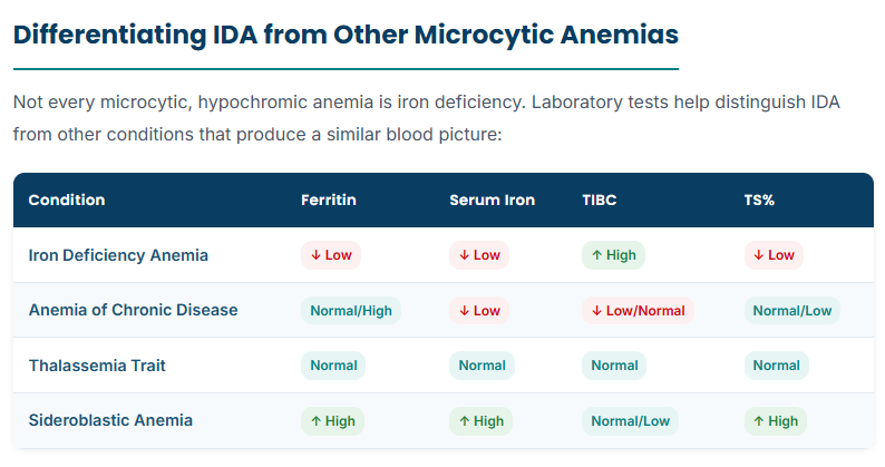
Quick Tip for Lab Professionals: When ferritin is in the low-normal range (12–30 ng/mL) but other iron markers suggest IDA, request a sTfR/log ferritin index. An elevated ratio strongly favors true iron deficiency over anemia of chronic disease.
Iron Deficiency Anemia Labs in Special Populations
Pregnant Women
Physiological hemodilution during pregnancy lowers hemoglobin naturally. WHO defines anemia in pregnancy as Hb below 11 g/dL. Ferritin below 30 ng/mL in a pregnant woman warrants iron supplementation even before anemia develops.
Children
Age-specific reference ranges apply to all CBC parameters in children. IDA is the leading cause of anemia in children under 5. A hemoglobin below 11 g/dL combined with low MCV and low ferritin is diagnostic in this age group.
Elderly Patients
In older adults, IDA is often caused by occult gastrointestinal blood loss. Laboratory investigation must be paired with clinical evaluation to find the underlying cause — new-onset IDA in an elderly patient should prompt gastrointestinal investigation to exclude malignancy.
Conclusion
Iron deficiency anemia is entirely diagnosable — and treatable — when the right laboratory tests are ordered and interpreted correctly. The complete picture requires more than a hemoglobin check. A systematic approach using iron deficiency anemia labs including CBC, serum ferritin, serum iron, TIBC, transferrin saturation, and peripheral blood film allows accurate diagnosis across all patient populations and disease stages.
For laboratory professionals: understanding the working principle behind each test improves quality control, reduces preanalytical errors, and enhances your ability to flag clinically significant results. For clinicians: integrating all iron panel values — not just a single number — leads to more confident, evidence-based management decisions.
Frequently Asked Questions (FAQ)
What is the most important lab test for iron deficiency anemia?
Can a normal CBC rule out iron deficiency anemia?
Why is TIBC high in iron deficiency anemia?
When iron stores are low, the liver increases production of transferrin — the iron-transport protein — to maximize iron capture from circulation. A higher transferrin level means more binding sites are available, raising the TIBC value.