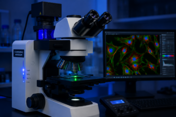

The bright field microscope is the most widely used microscope in laboratories, colleges, and hospitals around the world. If you are a medical laboratory technology student or a working lab technician, understanding the bright field microscope is one of the very first skills you must master.
This guide explains everything about the bright field microscope in simple language. You will learn its history, working principle, parts, types, uses, and how to take proper care of it. By the end, you will have a complete and practical understanding of this essential laboratory tool.
A Brief History of the Bright Field Microscope
The story of the bright field microscope begins in the late 16th century. Early lens makers in the Netherlands, including Zacharias Janssen, experimented with combining two lenses inside a tube to magnify small objects. This simple design became the foundation of the compound microscope.
In the 17th century, Antonie van Leeuwenhoek improved lens grinding techniques and became the first person to observe bacteria, red blood cells, and sperm cells using a simple single-lens device. His work opened the door to microbiology as a science.
Over the following centuries, instrument makers refined the optical and mechanical design. By the 19th century, companies such as Carl Zeiss and Ernst Abbe had introduced scientific principles of resolution and lens correction, turning the bright field microscope into the precise instrument we use today.
What Is a Bright Field Microscope?
A bright field microscope is an optical instrument that produces a magnified image of a specimen by passing ordinary light directly through it. The specimen appears dark or coloured against a bright background, which is where the name “bright field” comes from.
This type of microscope is the standard equipment found in almost every biology and pathology laboratory. It is affordable, easy to operate, and suitable for viewing stained smears, tissue sections, and many types of cells and microorganisms.
Working Principle of the Bright Field Microscope
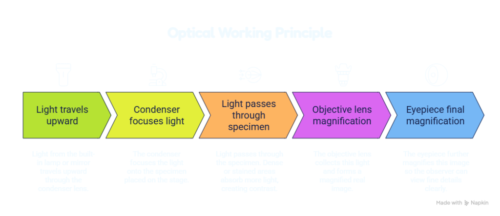
The working principle of a bright field microscope is based on simple transmitted light illumination and lens magnification. Understanding this principle helps you use the instrument correctly and troubleshoot common problems.
Optical Working Principle
- Light from the built-in lamp or mirror travels upward through the condenser lens.
- The condenser focuses the light onto the specimen placed on the stage.
- Light passes through the specimen. Dense or stained areas absorb more light, creating contrast.
- The objective lens collects this light and forms a magnified real image.
- The eyepiece further magnifies this image so the observer can view fine details clearly.
Mechanical Working Principle
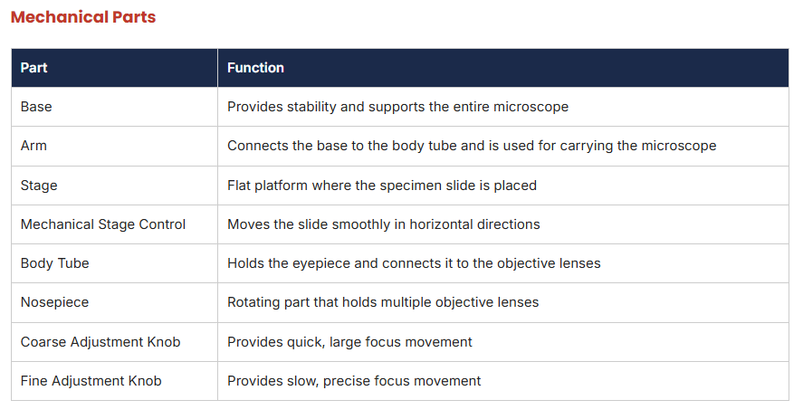
Alongside the optical path, the bright field microscope depends on precise mechanical movement. The coarse adjustment knob moves the stage or body tube quickly to bring the specimen roughly into focus. The fine adjustment knob then allows very small, controlled movements for sharp focusing. The mechanical stage lets the user move the slide smoothly in the X and Y directions without touching it directly.
Practical Tip: Always start focusing at the lowest objective power before switching to higher magnification. This protects the lens and the slide from accidental damage.
Classification and Types of Bright Field Microscope
The bright field microscope can be classified in a few different ways based on lens number, illumination source, and viewing style.
1. Based on Number of Lenses
- Simple Bright Field Microscope: Uses only a single magnifying lens. It offers limited magnification and is mostly used for basic observation or teaching purposes.
- Compound Bright Field Microscope: Uses two or more lens systems, an objective lens and an eyepiece, arranged to achieve high magnification with better resolution. This is the standard microscope used in modern laboratories.
2. Based on Viewing Style
- Monocular Bright Field Microscope: Has a single eyepiece for viewing with one eye. Common in schools and basic training labs.
- Binocular Bright Field Microscope: Has two eyepieces, allowing comfortable viewing with both eyes. Widely used in diagnostic laboratories.
- Trinocular Bright Field Microscope: Includes a third port for attaching a camera, useful for photomicrography, teaching, and documentation.
3. Based on Light Source
- Mirror-based Microscope: Uses natural or external light reflected through a mirror. Rare today but still found in some older teaching models.
- Electric or LED Microscope: Uses a built-in bulb or LED light source, offering consistent and adjustable brightness. This is the modern standard in hospitals and colleges.
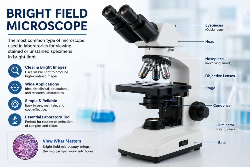
Main Parts of the Bright Field Microscope
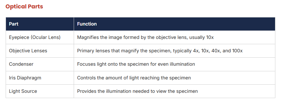
The bright field microscope is built from mechanical parts, which support and move the instrument, and optical parts, which are responsible for magnification and image formation.
Uses and Applications of the Bright Field Microscope
The bright field microscope has a wide range of uses across medical, educational, and research settings.
- Examining stained blood smears for cell morphology and blood-related disorders
- Identifying bacteria, parasites, and fungi in clinical samples
- Studying tissue sections in histopathology
- Teaching basic cell biology and microbiology in schools and colleges
- Routine urine and stool sample examination in diagnostic laboratories
- Quality checks in food, water, and pharmaceutical industries
Advantages and Limitations
Advantages
- Simple to operate and does not require complex training
- Affordable compared to advanced microscopy systems
- Widely available in laboratories worldwide
- Good for viewing stained specimens with clear contrast
Limitations
- Cannot provide good contrast for live, unstained, transparent specimens
- Resolution is limited compared to electron or phase contrast microscopy
- Requires proper staining technique for best results
Care and Maintenance of the Bright Field Microscope
Proper care extends the life of a bright field microscope and keeps image quality sharp. Laboratory technicians and students should follow these essential maintenance practices.
- Clean lenses only with lens paper or a soft microfiber cloth, never with regular tissue
- Use a small amount of xylene or lens cleaning solution to remove oil after using the 100x objective
- Always carry the microscope with one hand on the arm and one hand under the base
- Cover the microscope with a dust cover when not in use
- Store it in a dry cabinet away from moisture and direct sunlight
- Keep the stage and mechanical stage screws lightly lubricated as per the service schedule
- Switch off the light source immediately after use to increase bulb life
- Schedule periodic servicing with a qualified technician for calibration and alignment checks
Practical Tip: Never leave the 100x oil immersion objective in contact with a dry slide for a long time. Clean it right after use to avoid lens damage.
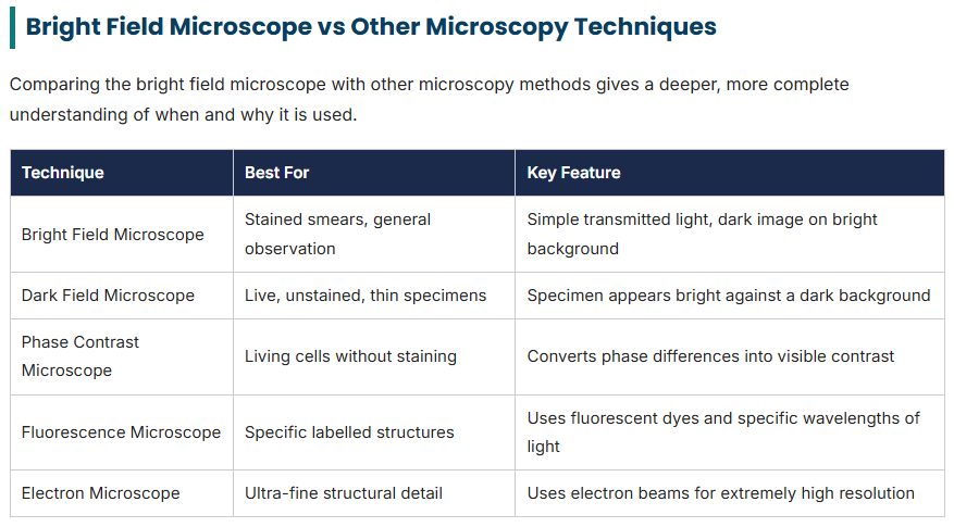
This comparison shows that while newer technologies offer more advanced imaging, the bright field microscope remains the reliable starting point for most laboratory training and diagnostic work.
Further Reading and Research Resources
For students who want to explore the scientific literature on microscopy and laboratory diagnostics in more depth, academic research databases are extremely useful. You can search peer-reviewed studies on PubMed and full-text articles on PubMed Central. Broader academic search across journals is also possible through Google Scholar.
Conclusion
The bright field microscope is a foundational tool in laboratory science. From its origins in the workshops of early lens makers to its role in modern hospitals and colleges, this instrument has stood the test of time. Learning its parts, working principle, types, and maintenance is a valuable and rewarding step for every medical laboratory technology student and professional.
Frequently Asked Questions
What is a bright field microscope used for?
A bright field microscope is used to examine stained and unstained specimens such as bacteria, blood cells, tissue sections, and parasites under normal transmitted light. It is the most common microscope used in medical laboratories, schools, and research centers.
What is the working principle of a bright field microscope?
A bright field microscope works by passing light from a light source through the specimen, condenser, objective lens, and eyepiece. The specimen appears dark against a bright, illuminated background because it absorbs some of the light passing through it.
What are the main parts of a bright field microscope?
The main parts include the base, arm, stage, body tube, eyepiece, objective lenses, condenser, iris diaphragm, coarse and fine adjustment knobs, and the light source.
A bright field microscope is used to examine stained and unstained specimens such as bacteria, blood cells, tissue sections, and parasites under normal transmitted light. It is the most common microscope used in medical laboratories, schools, and research centers.
A bright field microscope works by passing light from a light source through the specimen, condenser, objective lens, and eyepiece. The specimen appears dark against a bright, illuminated background because it absorbs some of the light passing through it.
The main parts include the base, arm, stage, body tube, eyepiece, objective lenses, condenser, iris diaphragm, coarse and fine adjustment knobs, and the light source.
What is the difference between simple and compound bright field microscopes?
A simple bright field microscope has only one lens for magnification, while a compound bright field microscope uses two or more lenses, an objective lens and an eyepiece, to achieve much higher magnification and clearer image resolution.
How do you maintain a bright field microscope properly?
Proper maintenance includes cleaning the lenses with lens paper and a mild cleaning solution, covering the microscope when not in use, storing it in a dry and dust-free place, avoiding rough handling, and servicing it regularly by a qualified technician.
A simple bright field microscope has only one lens for magnification, while a compound bright field microscope uses two or more lenses, an objective lens and an eyepiece, to achieve much higher magnification and clearer image resolution.
Proper maintenance includes cleaning the lenses with lens paper and a mild cleaning solution, covering the microscope when not in use, storing it in a dry and dust-free place, avoiding rough handling, and servicing it regularly by a qualified technician.